Our novel CPNs™ have been used in a wide range of life science research, including a study with precision instrument manufacturer, HORIBA. The outcomes of this work have demonstrated a novel method for the determination and identification of tumour margins.
The research makes use of the FLIMera, a new TCSPC based wide field Fluorescence Lifetime Imaging (FLIM) camera, to obtain real time fluorescence lifetime images from a tissue phantom containing a tumour mimic. The aim of the work is to see if fluorescence lifetimes, obtained using the very sensitive time-correlated single-photon counting technique, could be applied as a tool to distinguish tumour margins. This is important as the use of fluorescence guided surgery is increasing, especially in relation to cancer treatment. Fluorescent dyes are being employed to assist the surgeons in demarcating tumour boundaries.
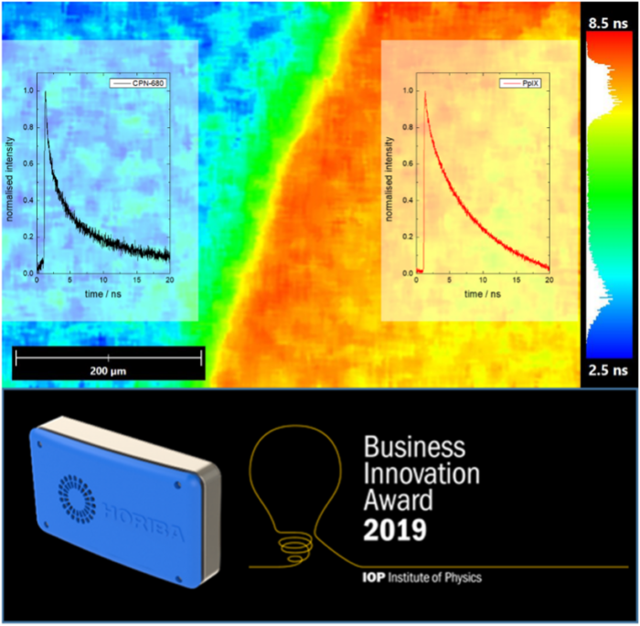
The article principally uses Protoporphyrin IX (PpIX) as the fluorophore. This mimics the action of 5-ALA, which can be given to patients and is metabolised in cancer cells to form the fluorescent PpIX. This fluorophore exhibits a large wavelength separation between where it is excited and emits fluorescence. To be able to show that fluorescence lifetimes can be used to distinguish between PpIX and a fluorescent background, CPN™ 680 was also incorporated within a tissue construct.
This dye was advantageous as it exhibited a similar wavelength shift to the PpIX, enabling both of the dyes to be excited at the same wavelength. Their emissions were monitored at the same time, but because of a difference in their lifetimes it was possible to image the PpIX in the tumour mimic and distinguish it from CPN™ 680 in the bulk of the phantom.
Conversation and collaboration between the two businesses came through two successful IOP Business Innovation Awards, with Stream Bio and HORIBA winning in 2018 and 2019, respectively.