In collaboration with the University of Leeds, we have been demonstrating the potential of CPNs™ in colon cancer cell imaging. With their powerful signal strength, CPNs™ can bind to and highlight cancerous cells in a patient’s body, enabling better resection of diseased tissue. CPNs™ could provide the sensitivity needed to ensure as much healthy tissue as possible is spared.
Carcinoembryonic antigen (CEA) acts as a biomarker for cancers, including colon cancer. CPNs™ can be combined with affimers – proteins designed to have a high affinity for specific biomarkers. A combined CPN and anti-CEA affimer nanoparticle can specifically bind to cells expressing CEA, highlighting where cancerous cells are present. Initial experiments show that CPN + anti-CEA affimers can effectively label cells expressing CEA and cell spheroids acting as tumour models.
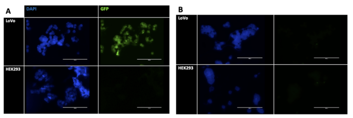
LoVo cells derived from a colorectal adenocarcinoma express CEA, while HEK293 cells do not. In Figure 1 A, DAPI highlights cell nuclei in both cell types, while CPN™ 510B combined with the targeting affimer only shows up in the cells expressing CEA. In Figure 1 B, when the CPN™ 510B is used without the targeting affimer, no green fluorescence is present. This shows that the fluorescent CPN 510B + anti-CEA affimer combination effectively marks the colon tumour cells.
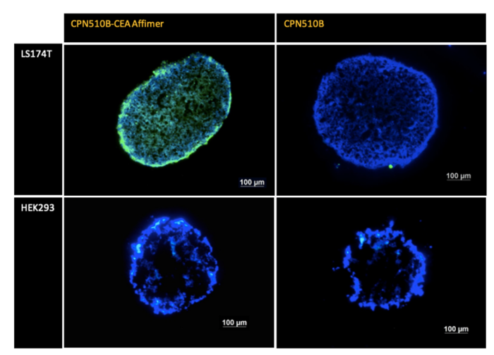
Figure 2 shows that this effect is still present in spheroid models, demonstrating that the CPN™ 510B anti-CEA affimer combination can penetrate tumour-like spheroids. This could have important implications for studying colon cancer tumour models and enhancing current fluorescence-guided surgery techniques.