Stream Bio has collaborated with a group at the National Physics Laboratory (NPL) to produce super-resolution images of Conjugated Polymer Nanoparticles (CPNs™) to better demonstrate their consistent size and brightness.
Super-resolution microscopy is a widely used technique, able to highly resolve structures in the nanoscale. This high resolution is not possible in conventional light microscopy due to the diffraction limit, but is achieved in super-resolution microscopy by modulating the excitation or activation light. The technique makes it possible to view individual molecules, allowing the study of sub-cellular structures and molecular interactions.
The team at NPL imaged CPNs™ using a Structure Illumination Microscope (SIM). After curing a CPN™ sample for 24 hours, the images captured display stable, high-intensity fluorescence throughout a range of wavelengths. Each CPN™ is clearly resolved, with a consistent diameter of approximately 70 nm.
As a result of their consistent size and fluorescence, CPNs™ are ideal for use as calibration beads in super-resolution microscopy. In addition, their magnetic core and propensity for conjugation with a variety of target molecules enable CPNs™ to enhance a broad range of cell biology studies.
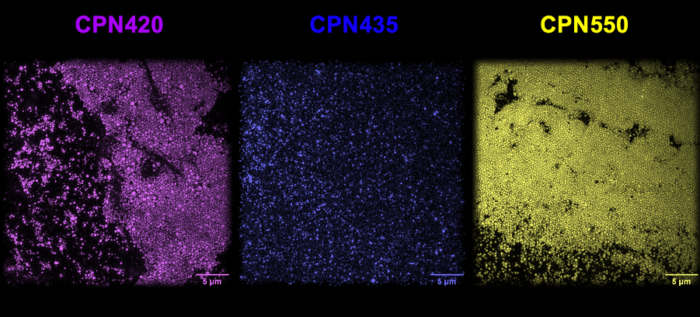
CPN™ 420 (Violet), CPN™ 435 (Indigo) and CPN™ 550 (Yellow) imaged using super-resolution microscopy at the National Physics Laboratory